| CHECK OUT OUR NEW EDUCATIONAL SECTION: DERMALSCAN NEWS |
 |
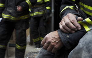
Screening Firefighters for Skin Cancer: FDNY Joint Initiative with Health Experts & Advocates
6/1/2024‐ FDNY Fire Academy (Randalls Island, NY). The FDNY united with the Firefighter Cancer Support Network (FCSN) and the American Academy of Dermatology (AAD) to conduct a comprehensive skin cancer screening for all of its active and retired members. Attendees appreciated the well‐orchestrated group effort in support of our first responders. Under the clinical expertise of Dr. Christine Kannler (dermatologist), she and her team installed a total of twelve examination tents at the ready for the many active and retired members of the FDNY. Thirteen board‐certified dermatologists and dermatology residents provided free skin checks to nearly 300 firefighters on Saturday. Dr. Kannler initiated her own version of the program since 2018, donating her valuable expertise and time to provide free, potentially life‐saving skin cancer screenings to firefighters under the AAD skin cancer screening program. |
|
|
|
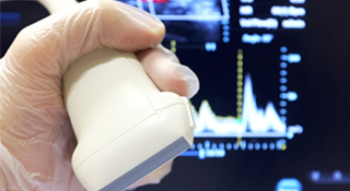
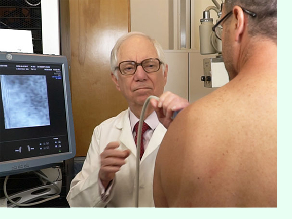
RCM PERFORMANCE TESTING WITH ULTRASOUND COMPLEMENT
On March 25, 2024, Mr. Steven Ridge (VP Sales & Marketing) and Mr. Wes Young (Clinical Applications Mgr) presented a full‐demo and contributed a 1‐day test drive to the IHRC Lab & Dr. Robert Bard's NYC office with select patients. They agreed to a full diagnostic performance review of the Vivascope 1500 while Dr. Bard mirrors the process with his ultrasound probe(s). As a supportive imaging device for dermatologists, medical leaders and researchers alike have embraced the RCM as the “go to” solution for complex disorders like skin cancers, alopecia, melanoma, burns, scars etc. Meanwhile, use of medical imaging solutions such as the ULTRASOUND, gives us that objective and quantifiable data that we need to monitor therapeutic progress and measure the current state of a patient’s pathology. Acquiring this form of data says, “this is where you were, this is where the treatment is, and this is where you are now!” (see full report)
|
SPOTLIGHT: DR. XIMENA WORTSMAN - A VISIONARY IN DERMATOLOGIC ULTRASOUND FOREWORD: By Dr. Robert L. Bard
To be part of an international and multidisciplinary society where we can witness true visionaries and pioneers lay the groundwork for the future of our science. It is a joy to meet and collaborate with clinical leaders and educators like Dr. Ximena Wortsman - a direct inspiration to my own commitment to exploring the many facets of ultrasound technology for both patient care and clinical research. Her campaign to make the concept of “dermatologic ultrasound” a globalized paradigm and a movement is nothing short of leadership at its finest. Dr. Wortsman’s textbooks, articles and lectures speak volumes for the future of integrative diagnostic medicine, forging a refreshingly modern view of dermatologic analysis, light years beyond convention. Her generous work directly supports the continuing education and the advancement of technical innovation in diagnostic and therapeutic medicine. It is for this reason that HealthTech Reporter recognizes the remarkable work of Dr. Ximena Wortsman. Her recent lectures at the 2024 UltraCon event highlighting her clinical expertise in ultrasound diagnostic imaging in dermatology are priceless resources for the medical community at large. (full article) |
|
 |
DERM EXPO NEWS Nov 30 – Dec. 3, 2023: The 26th Annual Mount Sinai Winter Symposium took place at The New York Academy of Medicine (NYC). This nationally recognized, live CME event was attended by over 550 participants. It offered a comprehensive program addressing Medical and Surgical Dermatology, which was delivered by 40 sought-after faculty speakers and world-renowned leaders in the treatment of various skin diseases and conditions. Topics included the most current research on the diagnosis and treatment of medical dermatological conditions, such as psoriasis, eczema, alopecia, hidradenitis supportive, actinic keratosis, rosacea, and skin cancer. In addition, cosmetic procedures were performed in front of the audience allowing them to witness the latest techniques in skin rejuvenation. |
|
|
WHAT IS PBM? (From MSK to Skin Disorders to Cancer Tumors)
Photobiomodulation (PBM) is a form of light therapy that uses red and near-infrared light sources to treat a variety of skin conditions. PBM is often administered using low-level lasers or light-emitting diodes, and has several advantages over other treatments, including being non-invasive, cost-effective, and easy to apply. PBM and phototherapy are becoming promising therapeutic approaches for treating a wide range of cutaneous diseases and disorders, including psoriasis, acne, atopic dermatitis, hair regrowth, wound healing, skin rejuvenation, and pigmentation disorders |
|
|
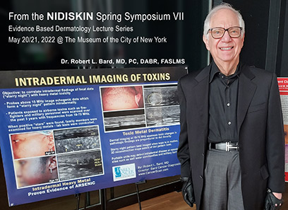
DERM-NEWS: TOXINS IN THE SKIN
According to the CDC, DERMAL ABSORPTION happens when a chemical goes through the skin and travels into the body. Many chemicals used in the workplace or even from home (ie. pesticides and organic solvents) can wreak havoc on and under the skin, damage internal organs and also the immune system if they penetrate the skin and enter the bloodstream. Most efforts to address chemical hazards have been focused on breathing, digesting or drinking chemicals rather than what's being absorbed through skin. Because of this, there are far fewer methods and campaigns dedicated to assessing skin exposures to toxins - UNTIL NOW.
With the latest evidence of inflammatory skin disease and foreign bodies under the skin, the medical and aesthetics communities are now facing the many new ways that environmental (toxic) influencers can affect the body through skin contact and absorption. This initiative is achieved thanks to our non-invasive diagnostic imaging advancements including Confocal Microscopy and the 3D Doppler Ultrasound. Dr. Robert Bard, presents this critical health topic as part of his 2022 Series on INTRADERMAL IMAGING OF TOXINS in dermatology conferences such as the NIDISKIN Spring Symposium. (See INFLAMMATORY SKIN DISEASE Poster)
|
|
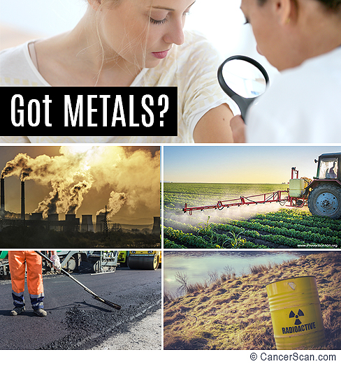
HEALTH ALERT: Skin Reactions Potentially Linked to Heavy Metal Contamination
Heavy metal poisoning can occur from a signifiant exposure to certain metals in the air, water or digested materials. Heavy metals including ARSENIC, LEAD and MERCURY are recognized to cause a wide range of health issues and even death and others. Poisoning can happen if you eat or drink
something tainted with heavy metals or if you breathe in contaminated dust or fumes.
ARSENIC EXPOSURES & CANCERS
Inorganic arsenic is a confirmed carcinogen and is the most significant chemical contaminant in
drinking‐water globally...and are highly toxic. According to the World Health Organization (WHO), people are
exposed to elevated levels of inorganic arsenic through drinking contaminated water, food preparation and
irrigation of food crops, industrial processes... and is used industrially in the processing of glass, pigments, textiles,
paper, metal adhesives, wood preservatives, ammunition, and, to a limited extent, in pesticides, feed additives
and pharmaceuticals.
The most common reason for long-term exposure is contaminated drinking water. Groundwater most often becomes contaminated naturally; however, contamination may also occur from mining or agriculture. It may also be found in the soil and air. Recommended levels in water are less than 10–50 µg/L (10–50 parts per billion). Other routes of exposure include toxic waste sites and traditional medicines. Most cases of poisoning are accidental. Arsenic acts by changing the functioning of around 200 enzymes. Diagnosis is by testing the urine, blood, or hair. [1][3][5]
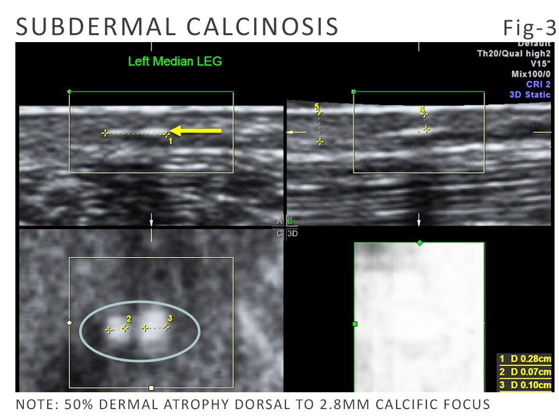
FIG-3 (R): This is a 3D image of subdermal calcification, which is obtained in 10 seconds over a four by four square centimeter area. In addition to showing how deep the calcium is (intradermal) as opposed to on the surface or below the surface and in the soft tissues, it shows intradermal calcium, the 3d aspect, quantifies, how many calcific areas are imageable in this picture.
Arsenic is a known human carcinogen associated with skin, lung, bladder, kidney, and liver cancer. A new study from the NTP Laboratory that replicates how humans are exposed to arsenic through their whole lifetime found that mice exposed to low concentrations of arsenic in drinking water developed lung cancer. The concentrations in the drinking water given to the mice were similar to what humans, who use water from contaminated wells, might consume. [NIH6]
PLEASE SEE THE COMPLETE UPDATED REPORT ON THE "GOT METALS" PROJECT. Imaging Identifies Subdermal Calcification & Exploring Links to Heavy Metal Toxicity
|
|
An Occupational Hazard
It is widely known that skin cancers or Melanoma comes from excessive tanning or high exposure to the sun. But reports have also shown other sources of Melanoma cases from work-related exposure to toxins. From 9/11 related accounts alone, conditions like Melanoma and Skin Cancers were early covered by the WTC Health Program due to identified cancer claims by first responders and volunteers from Ground Zero [1]. The CDC and a new study from the National Institute for Occupational Safety and Health (NIOSH) also examined an increase in firefighters aquiring skin cancers [2] and a Cancer Registry and found that firefighters had increased risks for several major cancers. Additional reviews from the NIH identifies connections between fighting fires and skin cancers. [3]
Also review how to avoid the risks of UNNECESSARY BIOPSIES |
COLLABORATIVE PROJECT: 2022: "IMAGE GUIDED AESTHETIC PROCEDURES & TREATMENTS"
Springer Medical Publishing proudly presents the first installment in clinical aesthetic procedures. This detailed and up-to-date overview of image-guided procedures focuses on the many aesthetic and reconstructive strategies delivered by some of today's renowned leaders in the clinical aesthetics community. They share their valuable expertise and field-based findings throughout this feature-rich textbook. The wide list of audiences for this text (ie. dermatologists, plastic surgeons, aestheticians, general surgeons) will enjoy an insider's look at each treatment program while providing remarkable field-based knowledge for the general non-medical audience seeking the latest information in non-invasive and minimally invasive aesthetic procedures.
Produced and edited by Dr. Robert L. Bard, (NYC based cancer diagnostic imaging specialist) this collective project showcases the most highly sought-after cosmetic treatments in each priceless chapter- through detailed breakdowns, experiential insights and a generous graphic tour of before and after progress visuals. Thanks to the additional safety benefits of clinical imaging, our treatment professionals express added confidence in the pre-operative and post-op areas. In addition, many aesthetics procedures noted also brings significant advantages (of accuracy and efficiency) to the actual treatment process from real-time image guidance. Some of our top contributors include: Dr. Beth Haney, Dr. Michelle Peters-Zappas, Mary Nielsen Aesthetics, Arun Garg, DMD and January Howard, CMA.
 . .
WHO IS DR. MICHELLE ZAPPAS?
Dr. Michelle (Shelly) Zappas is a family nurse practitioner and an aesthetics clinician at the Luxe Aesthetics Center as a clinician or as a family nurse practitioner in Yorba Linda, CA. In July of 2021, Dr. Zappas was recruited by Springer editor Dr. Robert Bard (NY Cancer Imaging Specialist) and co-editors at the NY Aesthetics Alliance to co-author a multi-disciplinary text project about IMAGE GUIDED AESTHETIC PROCEDURES. She and 12 others were tasked to produce comprehensive chapters on their specific expertise, making this the first multidisciplinary textbook of its kind. (See complete textbook overview) |
|
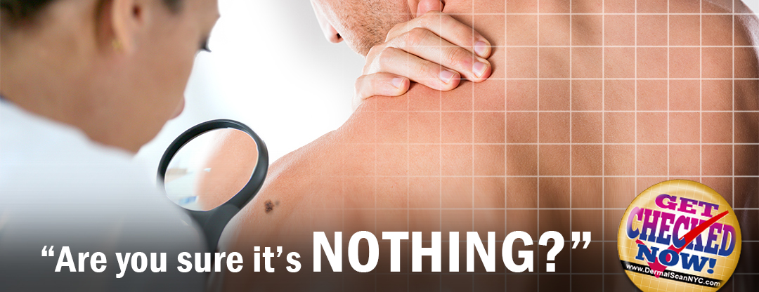
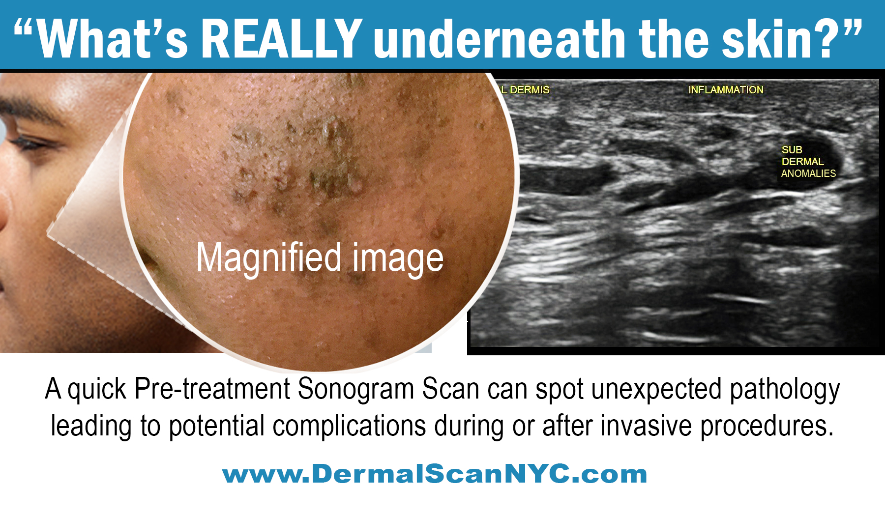
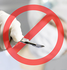
Avoid the risks of unnecessary BIOPSIES
When it comes to finding abnormalities in a patient exam, many conventional-minded doctors tend to tread on the side of caution... but usually at YOUR expense! They find an unusual spot that appears questionable and their first reaction is to cut it out and send it to the lab for a BIOPSY. As with all invasive surgical procedures (however large or small) carries risks including bleeding, infections, post-surgical scars and potential damage to nearby tissues and organs.
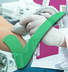
The year is 2019- the era of non-invasive technologies where identifying what's under the skin no longer needs to be about cutting into it. The age of robotics, artificial intelligence (AI), highly developed laser applications and advanced sonic diagnostic protocols are all fast replacing the age-old scalpel as part of risk reduction, time/cost advantages and increased performance in the world of clinical diagnostics and medical treatment.
Imaging technologies like the 4D Doppler Ultrasound™ can accurately and successfully scan, study and fully diagnose any skin anomalies and sharply view what's going on underneath. Derm imaging specialists stand on the side of innovation as they confidently rely on the most current devices to deliver the most accurate readings while bringing significant reduction to patient stress under a scan- that takes mere minutes!
Click here to review the latest diagnostic CancerScan technologies |
|
ENVIRONMENTAL RISKS ON OUR LARGEST ORGAN
According to the National Cancer Institute, SKIN CANCER is the most common type of cancer. The main types of skin cancer are squamous cell carcinoma, basal cell carcinoma, and melanoma. Melanoma is much less common than the other types but much more likely to invade nearby tissue and spread to other parts of the body. Most deaths from skin cancer are caused by melanoma. Explore the links on this page to learn more about skin cancer prevention, screening, treatment, statistics, research, clinical trials, and more.
When imaging detects a region of interest or suspicion, it can also be used to direct selective biopsies to obtain very small tissue samples for further laboratory analysis (pathology). The use of imaging together with pathology gives the most accurate information about the size, location and aggressiveness of any cancer thus identified.
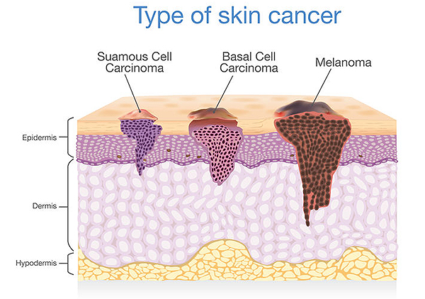 |
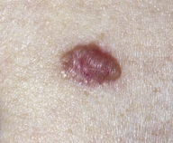
Skin Cancer
Non-melanoma skin cancer (NMSC) is the most common tumor in the adult population worldwide. While these are locally invasive and generally slow growing, some varieties (squamous cell carcinoma) may metastasize regionally and more widely. Skin cancer rates are continuing to rise and the most deadly form (melanoma) may be ruled out by 3D Doppler histogram imaging and advanced optical diagnostics. These lesions are often treated surgically, however, less invasive therapeutic options now exist allowing for better cosmetic outcomes. Premapping the depth and borders aids in treatment planning since the dermatologist recognizes disease that is visual and cannot see below the surface. The observation that the ear or nasal cartilage is involved changes the excision from a 5 minute procedure to a 5 hour operation. 3D imaging provides a roadmap for the surgeon and a patient guide to potential aggression and possible recurrence.
Actinic Keratoses: An AK forms when the skin is badly damaged by ultraviolet (UV) rays from the sun or indoor tanning. Most people get more than one AK. When you have more than one AK, you have actinic keratoses, or AKs. Anyone who has many AKs should be under a dermatologist’s care. Most people who have many AKs continue to get new AKs for life. AKs are considered precancerous. Left untreated, AKs may turn into a type of skin cancer called squamous cell carcinoma. (Source: AAD.org)
Basal cell carcinoma: Cancer that begins in the lower part of the epidermis (the outer layer of the skin). It may appear as a small white or flesh-colored bump that grows slowly and may bleed. Basal cell carcinomas are usually found on areas of the body exposed to the sun. Basal cell carcinomas rarely metastasize (spread) to other parts of the body. They are the most common form of skin cancer. Also called basal cell cancer.
Merkel cell carcinoma: A rare type of cancer that forms on or just beneath the skin, usually in parts of the body that have been exposed to the sun. It is most common in older people and in people with weakened immune systems. Also called Merkel cell cancer, neuroendocrine carcinoma of the skin, and trabecular cancer. (Source: NIH)
Squamous cell carcinoma: Cancer that begins in squamous cells. Squamous cells are thin, flat cells that look like fish scales, and are found in the tissue that forms the surface of the skin, the lining of the hollow organs of the body, and the lining of the respiratory and digestive tracts. Most cancers of the anus, cervix, head and neck, and vagina are squamous cell carcinomas. Also called epidermoid carcinoma. (Source: NIH)
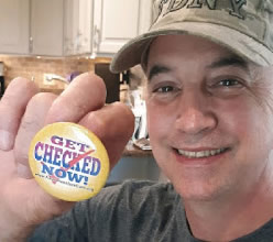
For complete information or to "GET CHECKED NOW!" visit: CancerScan.com |
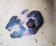
Melanoma
Malignant melanoma, one of the most lethal cancers, is increasing at an alarming rate. It is the leading cause of death in women aged 20-43 years. Importantly, only 1 out of 33,000 moles are malignant and sonography may prevent unnecessary biopsies. The chance of aggression is related to the tumor depth. Tumors less than 1 mm are often cured by biopsy. Disease greater in penetration is readily imaged by 3D Volumetric probes and non-palpable foci of tumor extension or lymph node (glands) involvement are visible in the scanned area. The metastatic potential is measured by the 3D Doppler study and follow up of distant tumor spread may be correlated with serial sonography. Some invasive surgical diagnostic procedures may be avoided by using high resolution imaging since scans detect tumor nests as small as 2 mm in the lymph nodes. This means that a sonogram guided needle biopsy may avoid the necessity of a massive radical operative dissection of otherwise healthy tissues. |
What is Get Checked Now?"
in the Spring of 2019, the NY Cancer Resource Alliance (NYCRA) united with a group of cancer organizations, advocates and educators to form a single message dedicated to early detection to battle cancer. This message addresses avoidable causes of cancer advancement which is due to proctrastination, denial or the lack of attention to regular checkups. A message to "stay vigilant and proactive" means staying constantly aware of one's health and how to prevent illness and chronic disease. To date, this remains the motto of all NYCRA partners and community organizations. |
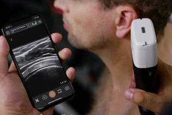
ACCIDENTAL BLESSING: “Pre-Op Nightmare" Averted by a HAND-HELD SCANNER
Based on a very true story | photo courtesy of www.Clarius.com
“After years of procrastinating, my husband Ted finally managed to book a trip to the dermatologist to remove a few annoying skin tags and a tiny mole from his left shoulder. On a routine trip to my radiologist, a simple handheld scanner concluded that Ted's mole was a MALIGNANT MELANOMA - a potentially deadly tumor. Ted discussed what would have been our next appointment and it was then that we realized that if the dermatologist would have applied the freezing solution to this mole under ‘standard procedure’ unaware of what we discovered, the melanoma would have metastasized and fast-tracked to every organ in the body.. . See Feature article |
|
MELANOMA IN THE NEWS
With our new environmental changes, skin-related issues are on the rise!Allure of the Summer Sun can be hazardous. Watch this feature coverage (CH11 WPIX) by Marvin Scott interviewing our very own Dr. Bard about the many risk factors that can cause Melanoma and Skin Cancer- and the preventative measures you can take to protect yourself.
|
|
PREVENTION & HEALTH CONSCIOUSNESS
According to the Surgeon General at the US Dept. of Heath & Human Services, the "seven Priorities are designed to improve health and wellness for the entire U.S. population, including those groups disproportionately affected by disease and injury."
• Avoiding risk factors and increasing protective factors may help prevent cancer.
• Being exposed to ultraviolet radiation is a risk factor for skin cancer.
• It is not known if the following lower the risk of nonmelanoma skin cancer:
• Sunscreen use and avoiding sun exposure
• Chemopreventive agents
• It is not known if the following lower the risk of melanoma: -
Sunscreen -
Counseling and protecting the skin from the sun
• Cancer prevention clinical trials are used to study ways to prevent cancer.
• New ways to prevent skin cancer are being studied in clinical trials.
(See complete Skin Cancer Prevention info @ the NIH website)
CANCER PREVENTION can be closely aligned with EARLY DETECTION. But from the perspective of a lifestyle upgrade, it is greatly proven that smart nutrition, toxin prevention (smoking, alcohol, drug abuse control), Stress management and Immune system support are all main ingredients to the prevention of cancers. These same protocols, for those who recently underwent cancer treatment, are what medical experts and wellness professionals prescribe to STAY IN REMISSION. |
EARLY DETECTION IMAGING PROGRAM
Historical reports show significant cancer claims from emergency rescue units- especially FIREFIGHTERS. This form of OCCUPATIONAL HAZARD dictates a high connection between long exposures to environmental toxins and cancer.
 An effective "weapon" in the battle against cancer includes Advanced Cancer Scanning to non-Invasive screenings. Dr. Robert Bard, expert Cancer diagnostician and the Northeast’s expert in 4D Doppler Imaging has developed the POST 9/11 CANCER DIAGNOSTICS PROGRAM. His NYC-based facilities (Bard Cancer Diagnostic Imaging) are equipped with only the most advanced state-of-the-art technologies that often out-performs higher-priced MRI’s, CT-Scan’s and X-Rays while promising to deliver more affordable and accurate reports in REAL-TIME from a process that's ready in MINUTES. An effective "weapon" in the battle against cancer includes Advanced Cancer Scanning to non-Invasive screenings. Dr. Robert Bard, expert Cancer diagnostician and the Northeast’s expert in 4D Doppler Imaging has developed the POST 9/11 CANCER DIAGNOSTICS PROGRAM. His NYC-based facilities (Bard Cancer Diagnostic Imaging) are equipped with only the most advanced state-of-the-art technologies that often out-performs higher-priced MRI’s, CT-Scan’s and X-Rays while promising to deliver more affordable and accurate reports in REAL-TIME from a process that's ready in MINUTES.
BARD CANCER DIAGNOSTIC IMAGING(NYC) has isolated and scanned countless cases of cancers using the most advanced diagnostic imaging technologies worldwide. We provide early detection and real-time "digital biopsies" of many tumor types using 4D Doppler innovations bringing accuracy and expediency to the comprehensive report- within MINUTES. For our patients, this is a priceless advantage that cuts down the wait time, decreases travel (to multiple diagnostic centers) and reduces the insurmountable level of stress and intolerable problems of today’s increasing medical bureaucracy. Our technology outperforms the advantages of MRI, X-ray and CT scans by 20-to-1. We have an uncompromising system that’s unique to the health care profession whereby our combined experience and technical advancements are called upon by many university hospitals and private practices today. For more information, contact Bard Cancer Diagnostics at 212.355.7017 or visit www.cancerscan.com |
|
| MODERN & ADVANCED DIAGNOSTIC SOLUTIONS |
|
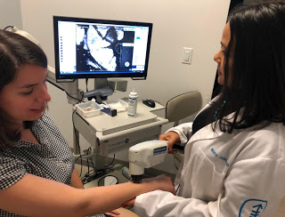
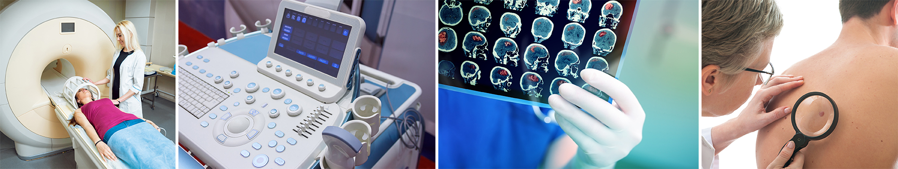
THE 3D/4D DOPPLER ULTRASOUND FOR ADVANCED DIAGNOSTIC IMAGING
For first responders, a NYC-based imaging facility Using state-of-the-art, high-speed equipment that's captures advanced cancer scans and subdermal screening in real-time through the use of COMPUTERIZED 3D DOPPLER HISTOGRAM technology. This innovation uses unique forms of sonography to evaluate blood flow related to tumor activity and to identify areas of suspicion. More tumor vessels signifies more aggressive disease. 3D analysis is non-invasive and rapid with results available to the patient during the visit. Patients in need of reassurance and world class imaging come from all countries for cancers of the prostate, breast, skin, thyroid and melanoma. For more info, visit: www.CancerScan.com
REFLECTANCE CONFOCAL MICROSCOPY-
THE LATEST IMAGING ADVANCEMENT FOR DERMATOLOGISTS
The modern era of diagnostic clinical imaging continues to expand in areas of optimal speed, sensitivity and feasibility as part of its continued pursuits to bring a non-invasive diagnostic modalities to our treatment community. The Reflectance Confocal Microscopy (RCM) gives dermatologists a major upgrade (over age-old microscopy) in their ability to assess pathologic and physiologic conditions of the skin with a higher level of clinical accuracy, greatly supporting the reduction of calls for biopsies of benign lesions. Responding to the limitations of biopsies and conventional screening methods, the non-invasive movement brings a heightened level of performance and responsiveness in areas of resolution, magnification, depth, contrast, and immediate results from bedside. Please see Dr. Manu Jain's complete technology tour @ Modern Diagnostic Science
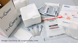
PART 3: THE LAB KIT - PUBLICLY AFFORDABLE & ACCESSIBLE
A recent trend in testing kits commercially distributed to physicians and/or direct to the consumer, are changing the way patients are receiving treatment and billed. Packaging lab testing services from anywhere in the country through a portable (mailable) transport kit is a most cost effective and efficient way to expand public access to and from any specialized lab service(s). One of the more prominent blood test kits available is called IVYGENE- allowing access to a specialized lab from anywhere in the country to partner with any clinic or practice. This streamlines the retrieval and delivery of acquired blood samples to the designated lab and greatly eliminating a number of steps and "overhead" expenses. Instead of building a high-priced lab in each city, county or state, there is now just once central lab for the entire country. For more info, visit our IVYGENE REPORT.
IVYGENE- CANCER DETECTION FOR FIREFIGHTERS
A firefighter's emergency rescue work often brings exposure to a wide range of hazardous (and potentially carcinogenic) substances. A recent study from the IAFF shows a significant percentage of firefighters will be diagnosed with cancer at some point during their lifetime. Others studies indicate that firefighters are up to 2X at risk for developing certain cancers. It is for this reason that First Responders HealthScan and its clinical partners including Integrative Medicine of NY helped to publicly introduce genetic testing protocols and such scanning/diagnostic innovations as the IvyGene solution to the community. Originally founded for 9/11 survivors and victims, the 911CancerScan program brings informational and resource access to any and all first responders to address this health risk. See the First Responders Cancer & Health Support Resource website for more information.https://healthscannyc.org/ |
|
EMPOWER YOUR TREATMENT PLANS WITH AN INDEPENDENT
SECOND OPINION
Knowing that you have CANCER can impair your ability to make the best choices. Gain a different outlook (with possible results) with a fresh pair of eyes and tools. Get new insights on your current health & lab reports and receive an alternate diagnostic imaging exam of your target area(s) to get validation or competing opinions on what’s truly going on inside. Confirm your primary diagnosis or possibly uncover false-positives and other misreadings that could change your entire treatment strategy altogether!
From the moment you receive the news about having cancer, you may grow curious about what other treatment options another oncologist would prescribe? You may want to get another doctor’s perspective on your current diagnosis or get another set of professional eyes to review your reports. All these concerns are quite natural and common - especially when it comes to pursuing such a major commitment as cancer care. We all want to gather all the information we can from trusted sources to build an intelligent direction that gives us complete confidence. That’s what a SECOND OPINION offers.
A Second Opinion helps affirm (or challenges) your current treatment strategy for your exact type of cancer. Finding another specialist outside of the prior doctor's circle of influence helps bring a different perspective to identifying any unique anomalies that other doctors and technologies may overlook. A second opinion is all about getting PEACE OF MIND in troubleshooting or problem solving your most vital health concerns
|
SELF-AWARENESS IS JOB 1!
This program is a simple plan to keep cancer away- or increase the likelihood of beating cancer with the comprehensive Early Detection & Prevention plan. In each case, much can be done to prevent the current stage. The first step is to GET MORE FAMILIAR WITH YOURSELF.
• Be aware of your genetic lineage: risk of cancer increases upon heredity. The first place to look is within your own dna or family history. Many cancers tend to travel down generations. It can also have the tendency to skip one generation and appear in the next one.
• Periodic Checking of your body for any anomalies like lumps, bumps, discolored bruises or growths. Self-checking is the first base. Also stay on top of unusual feelings like frequent headaches, unique pains and strains- anything that feels out of the ordinary. Take nothing for granted when it comes to your body.
• Know your environment: Many health issues are known to be caused by environmental toxins. Where you sleep, eat and work could be affecting how you feel later. Some health hazards are fairly visible and apparent while others may need some historical research in your area where there may have been potential chemical wastes or spills in the past. If you know of such issues, further research, demographic studies, protective measures and targeted checkups may be your next step.
ADDITIONAL LINKS: IDENTIFICATION OF WEAKENED IMMUNE SYSTEM AND HOW TO BOOST IT
|
|
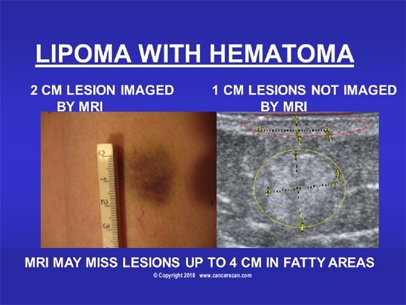 |
9/11 CANCER SCARE-SAME PATIENT 6 MONTHS AFTER TREATMENT
Our mesothelioma patient (pictured above) had a minor injury and noticed a new lump under the small bruise. A 5 minute scan showed the discoloration was caused by minor blood vessel damage due to the underlying BENIGN fatty tumor. Cysts and other non-malignant disorders can be distinguished from metastatic lymph nodes (glands) providing “real-time” relief of anxiety of cancer recurrence. |
|
|
ROBERT L. BARD, MD, PC, DABR, FASLMS- ADVANCED CANCER IMAGING SPECIALIST
Having paved the way for the study of various cancers both clinically and academically, Dr. Robert Bard co-founded the 9/11 Cancer Scan program to bring additional diagnostic support to all first responders from Ground Zero. His main practice in midtown, NYC (Bard Diagnostic Imaging- www.CancerScan.com) uses the latest in digital Imaging technology has been also used to help guide biopsies and in many cases, even replicate much of the same reports of a clinical invasive biopsy. Imaging solutions such as high-powered Sonograms, Spectral Doppler, sonofluoroscopy, 3D/4D Image Reconstruction and the Spectral Doppler are safe, noninvasive, and does not use ionizing radiation. It is used as a complement to find anomalies and help diagnose the causes of pain, swelling and infection in the body’s internal organs while allowing the diagnostician the ability to zoom and ‘travel’ deep into the body for maximum exploration.
 |
JESSE A. STOFF, MD, MDH, FAAFP- INTEGRATIVE ONCOIMMUNOLOGIST
Dr. Stoff is a highly-credentialed medical expert studying all medical remedies in pursuit of resolving the most challenging health issues of our time. In many circles, he is recognized for his 35+ years of dedicated work in immunology and advanced clinical research in modern CANCER treatments. He has spoken worldwide in some of the most sought-after medical conferences about his experiences and analyses on the study of human disease. His integrative practice INTEGRATIVE MEDICINE OF NY, Westbury, NY) has been continually providing all patients with the many comprehensive clinical options and modalities available- including "ONCO-IMMUNOLOGY", the science of battling cancer cells and reversing pre-cancerous conditions through a complete prevention program that has earned him great success in this field.Dr. Stoff has treated and managed countless patients who were affected by 9/11-related disorders and continues both public awareness efforts and clinical research in supporing this community of victims.
 |
CHERI AMBROSE, Co-editor / outreach coordinator for NYCRA
Cheri is the associate editor for various publications such as PinkSmart News, the Journal for Modern Healing and First Responders Cancer News. She is a patient advocate for many cancer-related programs and often contributes her time in cancer research fundraising events. As the communications director for the NY Cancer Resource Alliance, she manages community outreach, partnership missions with other cancer foundations and research organizations and attends educational functions for cancer awareness. Her latest public projects include the launch of ImmunologyFirst.org and ImplantScan.org. She stands as the current President of the male Breast Cancer Coalition (MaleBreastCancerCoalition.org).
|
| Disclaimer & Copyright Notice: Unless otherwise indicated on this Internet web site, you may display, download, archive, and print a single copy of any information on this Internet web site, or otherwise distributed from us for such personal, non-commercial use. Also, certain content may be licensed from third-parties. The licenses for some of this Content may contain additional terms. When such Content licenses contain additional terms, we will make these terms available to you on those pages, in the Terms of Use or in the Additional Information section of our Site (which his incorporated herein by reference). This website does not support, endorse or recommend any specific products, tests, physicians, procedures, treatment opinions or other information that may be mentioned on this site. Referencing any content or information seen or published in this website or shared by other visitors of this website is solely at your own risk. The publishers/producers of this Internet web site reserves the right, at its sole discretion, to modify, disable access to, or discontinue, temporarily or permanently, all or any part of this Internet web site or any information contained thereon without liability or notice to you. |
|