Nurse DEBI CAVOLO shares some of the top health disorders in women based on her research and professional experience!
"Putting ourselves first is not a luxury- but a health and wellness necessity!" states DR. ARTEMIS MORRIS, Naturopathic Physician.
DR. OSAMA KANDALAFT addresses the evolution in the medical community about underserved communities (including women).
Why support a Diagnostic-Based Network?
Though our professional group is certainly about a wide range of health resources and the collaboration of all types of professionals working on women's issues, a DIAGNOSTIC-based collective ensures a patient-dedicated road map and a more thoughtful health analysis and research-based evaluation. It is this commitment to a deeper sense of examination of the patient that lends itself to a more holistic and integrative strategy to therapeutics. It is also this philosophy that draws more intuitive and insightful clinicians to collaborate and share insights on a patient's disorder with
compassion and genuine care. (See "The Future in Personalized Medicine")
THE FIRST TRULY INTEGRATIVE PROGRAM FOR WOMEN’S HEALTH!
For over 35 years, Bard Diagnostic Imaging has been providing the most trusted health monitoring services for an expanded list of critical issues and concerns- with the latest in NON-INVASIVE, real-time imaging solutions. This year, we have united with other clinical experts to cover an even wider set of supportive services for women in areas of Prevention and Early Detection screenings alongside a comprehensive analysis of major Chronic Health Disorders.
An Alliance of Caring Experts Having access to the latest in compassionate experts and innovative diagnostic solutions for women is the first step to maintaining good health. Our alliance unites collaborative minds about the latest solutions from a wide range of modalities to offer optimum choices for all patients. Together, we provide expansive research and info-sharing about all clinical options and protocols as well as testimonials and clinical viewpoints from active professionals in the field.
Our Women’s Diagnostic Network is a collective partnership of independent clinical experts and medical researchers in specialized areas of study including CANCER CARE, ONCOLOGY, GYNECOLOGY, PHYSIATRY, DERMATOLOGY, NEUROLOGY, CARDIOLOGY & MENTAL HEALTH. Through medical collaboration, we unite to support improved patient evaluation, reporting and overall diagnostic care. For more information, contact us today at: 212.355-7017
Heart Disease is the #1 Killer of Women
According to the CDC, despite an increase in awareness over the past decades, only about half (56%) of women recognize that heart disease is their number 1 killer. Heart disease is the leading cause of death for women in the United States, killing 301,280 women in 2019—or about 1 in every 5 female deaths. We unite a network of clinical researchers, women's advocates and support groups to promote educational awareness about this national health alert.
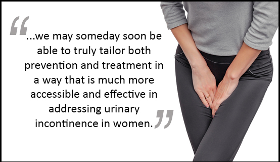
Innovations in Stress Incontinence:
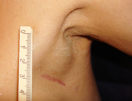
Anywhere from 26-40% of women are recorded to suffer from incontinence. Therapeutic experts and innovators find that the vast majority not know that there are treatment options and they resign themselves to a life of pads or diapers (and emotional challenges). A united alliance of global experts share valuable information about the current state of solutions about the various types of incontinence- including postpartum/stress incontinence. (see our feature article)
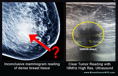
Reducing False Negative Readings of Cancer-Linked Dense Breasts
The medical community is now recognizing the link between dense breast tissue to the risk of breast cancer. This tissue type shows as a white cloud on a mammogram, cloaking a white tumor on a mammogram. As mammography prevails as the standard early detection screening test for breast cancer, the need for SUPPLEMENTAL SCANNING (with devices like an ultrasound) offers supportive detection of potential tumors that may be concealed by dense breast tissue under a mammogram. (see our feature article)
Diagnosing Female Incontinence through Imaging (4.5min)
A diagnosis is defined as the process of accurately and quantitatively determining health status, disorder or a diseased condition. One of various diagnostic options in modern medical science is through the use of imaging. Though this video narrated by Dr. Bard (resident imaging interpretation expert), we are able to visualize and better understand the physiology of Stress Incontinence- a popular postpartum disorder in women.
Editorial Feature: UNDERDIAGNOSED WOMEN (4.5min)
Historically, there is substantial evidence of gross disregard and dismissive response on the part of the medical establishment in the way women have been diagnosed. Critics have speculated everything from professional carelessness to blatant sexism (and other forms of discrimination) to a lack of updated education in current research. This compelling report starts with the landmark story of the legendary comedienne, Gilda Radner who died of ovarian cancer remission in 1989, then to the treatment complications of Nancy Cappello whose false negative mammography reports led to a fatal, late stage breast cancer(2018). Their stories remain as life-saving references about early detection. (see complete article)
What can Pelvic Floor Ultrasound do for the clinician?
Ultrasound has been used in the investigation of pelvic floor disorders since the 1980’s, not that long after its introduction into other gynaecological subspecialties such as Reproductive Endocrinology and Gyn. Oncology.
Progress in our field however has been much slower, and this deserves comment. Delays in the uptake of the new modality were partly due to the relative immaturity of urogynecology as a subspecialty, partly due to the fundamentally different way in which ultrasound was utilised in clinical practice.
The WOMEN'S DIAGNOSTIC NETWORK proudly offers 1 FREE DOWNLOAD/access to Dr. Hans Peter Dietz's PELVIC FLOOR ULTRASOUND: Atlas & Textbook. First published in 2016 by HP Dietz MD, PhD FRANZCOG DDU CU (Professor of Obstetrics and Gynaeoclogy/NSW Australia) - click to access
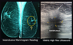
Ultrasound Significantly Reduces False Readings of DENSE BREASTS
7/8/2021- A wave of recognized medical sites, journals and reports are now indicating that dense breast tissue increases the risk of developing breast cancer and often masks a tumor from being seen on the mammogram since dense tissue is white and cancerous tissue is also white. Mammograms are the standard screening test for breast cancer, however, in the 21st Century, ultrasound non invasive imaging is the preferred exam for dense “lumpy” mammary disease.
[L- PLAY VIDEO] Dr. Nancy Cappello's Story: "I have dense breast tissue – and women like me (2/3 of pre-menopausal and 1/4 of post menopausal) have less than a 48% chance of having breast cancer detected by a mammogram. In November 2003 I had my yearly mammogram and my "Happy Gram" report that I received stated that my mammogram was "NORMAL" and that there were "no significant findings." Six weeks later at my annual exam... the mammogram revealed "nothing" yet the ultrasound detected a large 2.5 cm suspicious lesion, which was later confirmed to be stage 3c breast cancer, as the cancer had metastasized to 13 lymph nodes..." (click to see complete story)
TAMOXIFEN vs. AROMATASE INHIBITORS FOR MALE BREAST CANCER
In a private conference hosted by the Male Breast Cancer Coalition, our NYCRANEWS editorial team met Dr. Jose Pablo Leone, medical oncologist and researcher at the Dana-Farber Cancer Institute. Discussions covered tamoxifen and aromatase inhibitors for the treatment of male breast cancer, and his research plans in this field.
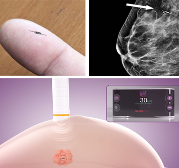
SURGICAL EVOLUTION IN BREAST CANCER LOCALIZATION:
WIRE-FREE RADAR IMPLANTS
Wire-free Radar Localization is a pre-surgical procedure to locate and mark the exact breast abnormality through the use of a small, 12×1.6 mm implanted radar reflector device, roughly the size of a grain of rice. This micro-electronic implant communicates with the scanning handpiece, allowing the surgeon to identify the exact tissue (and how much of it) to extract during a lumpectomy [1]. In this feature article, we present Dr. TroyShell-Masouras of Paradise Coast Breast Specialists in Naples Fla. - and David Gilstrap, Director & Global Product Management of Merit Medical. Together, we explored technical perspectives and design strategies behind radar localization and the SCOUT® technology. They shared the procedural advantages provided by the wire-free upgrade as well as its overall improvements to the patient's well-being in the pre and post-surgical phases. (See full article)
CLINICAL IMAGING OF BREAST CANCER Explained (click thumbnails for enlarged slide show)
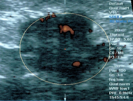
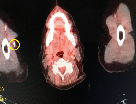
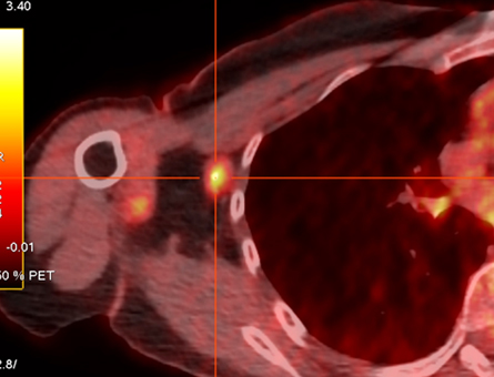
These images are an example of practical non-invasive imaging technologies that are currently used worldwide. The Doppler blood flow has been used for 30 years in Europe and Japan. Elastography (also non-invasive modality to show how hard or malignant a tumor is) has been around for 15 years with great success in many countries. A benign looking elastogram scan avoids biopsies in the thyroid, breast, prostate and lymph nodes.
While elastography is relatively new in the United States, the addition of Pet/CT with digital analysis is adding further specificity because chemotherapy, filler and benign tumors can show up as positive findings on a pet CT scan- we have to make sure that what's showing up as a bright lesion is not a false positive. The addition of documenting a hot area on the isotope Pet/CT scan is important to avoid false positives. The latest digital Pet/CT scans are more accurate and quantify true positives and allow you to avoid biopsying false positives. The orchestra of multiple complimentary non-invasive imaging technologies assures a quick and accurate way of determining cancer aggression and accurately allowing you to adjust treatment as needed in a timely matter.
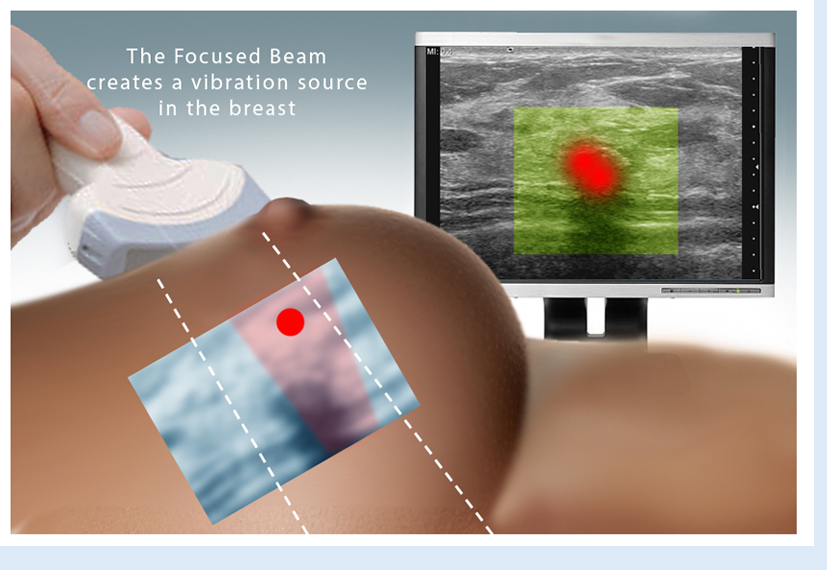
Three Staging Options for Advanced Digital Breast Imaging Advanced sonogram technology provides accurate use of sound waves to produce real-time images of the inside of the body. It is used as a complement to find anomalies and help diagnose the causes of pain, swelling and infection in the body’s internal organs while allowing the diagnostician the ability to zoom and ‘travel’ deep into the body for maximum exploration. Digital Imaging technology is also used to help guide biopsies and in many cases, even replicate much of the same reports of a clinical invasive biopsy. Imaging solutions such as high-powered Sonograms, Spectral Doppler, sonofluoroscopy, 3D/4D Image Reconstruction and the Spectral Doppler are safe, noninvasive, and does not use ionizing radiation. They also have the ability to provide early detection scans. ** Three STAGING OPTIONS are available depending on the patient’s current needs- to be discussed with practitioner prior to or during examination.
Option 1
Option 2
Option 3
BREAST only
Typically performed for patients either self-identifying anomalies (bumps) for the first time and/or are seeking a second opinion + may have a family history of breast cancer (or other related cancers related to the breast)
BREAST & AXILLAE
This option scans the breast and the shoulder through which vessels and nerves enter and leave the upper arm; a person's armpit. This option is recommended for anyone who may be currently Undergoing cancer treatment... also anyone who has recently (prior to 5 years) undergone a mastectomy since it detects unsuspected metastatic disease
BREAST, DUCTS &
LYMPH NODES
For a patient who is at high risk for developing a cancer or requires advanced confirmation of a current tumor treatment protocol since the 3D/4D computer analysis verifies the success of the therapy in a quantitative analysis by the unique Doppler histogram technology
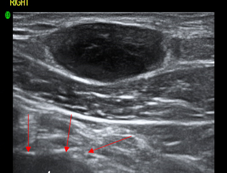
• SONOGRAM: Seek subdermal masses, breast lumps, tumor growths, fluid-filled cysts, nodes or hematoma. Sonography can be performed in multiple scan planes with real time 3-D image analysis with power and spectral Doppler flows.
• SONOFLUOROSCOPY: of intra/subdermal soft tissues is to be performed in multiple scan planes with varying transducer frequencies.
• 3D/4D IMAGE RECONSTRUCTION: For any lesions that are homogeneous echogenic and well marginated on 3D and 4D computer histogram analysis.
• SPECTRAL DOPPLER: Search for hyperemia of any lesions. Power Doppler will show normal flows in the adjacent arteries/veins. Spectral flows will show normal tri-phasic waveforms. Search for any peripheral lesion abnormalities or focal vascularity.
For more information on Advanced Digital Breast Imaging, contact us at: 212.355.7017
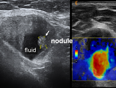
BREAST CANCER SCREENING REGIMEN The current standard in screening for breast cancer is mammography. However, this imaging tool misses some breast tumors, especially in women with dense breasts. Published data suggests that sonography can play an important role in detecting tumors that mammography misses. In fact, over 94% of cancers seen only on ultrasound were invasive tumors with average size of 10 mm, and in the studies where staging was detailed, 91% were node negative, meaning it had not spread and complete cure was possible due to early detection.
Power Doppler Sonography adds increased accuracy to breast imaging evaluation over ordinary ultrasound because it shows higher blood flow speeds, often a sign of cancerous activity in the breast. Studies have shown that suspicious blood flow identified by pre-surgery Power Doppler scans corresponds very well with the size, location and aggression of actual tumors that are then surgically removed. Thus, Power Doppler brings an important clinical dimension to breast cancer detection. 3D sonography clearly shows tumor margins and 3D Doppler affords an index of cancer aggression and metastatic potential.
Breast Sonograms and Ultrasound offers the following advantages:
• Highest accuracy in dense (lumpy, cystic) breasts
• Non-invasive-no radiation exposure
• Cost effective
• Can distinguish cysts (fluid-filled masses) from cancerous tumors without needle sampling
• Ease of image guidance for breast biopsy
Women who should consider ultrasound scanning of the breast include those at risk of breast cancer because of personal or family history and the presence of fibrocystic (dense) breast tissue which increases cancer risk by 400%.
Breast Imaging & Advanced Screening Programs
for New York Women's Groups & Organizations
LET'S UNITE IN THE FIGHT! In the spring of 2018, Bard Cancer Diagnostics (BCD) recently partnered with AWARENESS FOR A CURE and The NY Cancer Resource Alliance to join the battle against breast cancer by establishing a foundation-based Cancer Screening & Monitoring program for all members of non-profit Breast Cancer Organizations and NY-based Women's Groups. This unique program works with all major health providers to bring the most up-to-date technologies and alternative diagnostic solutions (including "2nd Opinion" reviews) and regularly scheduled monitoring and EARLY DETECTION SCANS - all at a significantly reduced rates. BCD also raffles off an Advanced Comprehensive 3/D & 4/D Diagnostic Packages for Breast, Skin and Lung Cancers for qualified organizations- a $2200 value. Inquire about our programs for Women's orgs: email bardcancercenter1@gmail.com
or call 631-920-5757. (vid1)
FAQ about Non-Invasive Sonic Technology & the Future of Cancer Biopsies
Since 1973, Dr. Bard established global recognition in the medical field through his contributions in the advancement of cancer diagnostic innovations. His special use of advanced imaging technologies are widely praised as the painless alternative and a more accurate innovation as well as a much faster solution for acquiring results over surgical biopsies. For the patient, it's a world of difference and immediate peace of mind when the 3D imaging establishes the diagnosis during the examination as you literally see the pictures in front of you in real time.
Q: How accurate is Advanced Sonography in identifying cancers?(open)
Q: Give us an example of your 4D Scan's accuracy over conventional diagnostic methods?(open)
Q: What types of cancers are most commonly captured with this technology?(open)
Q: How can digital scanning be instrumental in Early Detection?(open)
Q: What are the benefits of using the Doppler Ultrasound imaging for BREAST CANCER?(open)
Q: What are the most recent upgrades in the cancer imaging industry?(open)
Q: Can this technology be useful in other health issues and disorders?(open)
MPR-TV reports on Advanced Cancer Doppler Imaging. Meet Dr. Robert L. Bard (award-winning cancer diagnostician and expert imaging specialist) and see him break down the difference between a benign tumor and a fully-blown cancerous cell- and how using his advanced 3D and 4D Doppler technology makes all the time-saving difference in getting an accurate study.
SELF-AWARENESS IS JOB 1!
Many routine imaging procedures can assure people that they are at risk for a disease or catastrophic medical event: heart scans, virtual colonoscopies, and lung screening are a few examples of ways to encourage healthy choices (nutrition, supplements, exercise, stress management, etc.) by reinforcing them. BARD CANCER DIAGNOSTICS is founded on the commitment to explore and implement the latest diagnostic technologies as a means of building the proper treatment strategy of many types of cancers. We also specialize in PREVENTION solutions for our patients who strive to maintain a health-conscious lifestyle as well as those who are at increased risk of certain diseases (hereditary factors or environmental exposure to toxic substances) by confirming that their efforts to prevent disease are working.
COMPUTERIZED 3D DOPPLER HISTOGRAM ANALYSIS
Using state-of-the-art equipment, Dr. Bard’s practice offers unique forms of sonography to evaluate blood flow related to tumor activity and to identify areas of suspicion. More tumor vessels signifies more aggressive disease. 3D & 4D analyses are non-invasive and rapid with results available to the patient during the visit. Patients in need of reassurance and world class imaging come from all countries for cancers of the prostate, breast, skin, thyroid and melanoma.
PINK-SMART NEWS IS COMING BACK! In April of 2021, we are publishing new interviews, articles, videos and other educational must-haves for survivors, patients and the community that is breast cancer. With support from some of the top leading foundations, Pink-Smart News will be presented by some of the who's who in Oncology, Immunology, cancer radiology and other areas that support the advancement of cancer care. See archived articles from Pink-Smart News
ABOUT OUR PROGRAM
As of Jan '18, Dr. Robert Bard spearheaded a partnership with a host of cancer educators, medical practitioners and non-profit foundations (allied under AwarenessforaCure.org) to form a public resource program
to aid in the advancement of the public's understanding about self-preservation
from cancer and other chronic diseases. EARLY DETECTION & PREVENTION is a global health movement that promotes a higher regard for "clean living" - from toxins and a toxic lifestyle. Our program consists of four main efforts: EDUCATION, COMMUNITY CONNECTION, CURRENT NEWS & CLINICAL RESOURCES. EARLY DETECTION & PREVENTION brings the empowerment of wellness through group seminars, videos
and the distribution of current articles & newsletters published/shared to all the major cancer charities and their
members.
For more information or to subscribe to our EARLY CANCER DETECTION & PREVENTION PROGRAM newsletter, contact Bard Cancer Diagnostics
today at: 212.355.7017 - or email us at: bardcancercenter1@gmail.com. Bard Cancer Diagnostics is located at 121 E. 60th St. Suite #6A New York, NY 10022. Visit the complete website: www.CancerScan.com
EARLY DETECTION SAVES LIVES! According to the World Health Organization, early detection of cancer greatly increases the chances for successful treatment. In the ongoing battle against cancer, it is common knowledge that most cancers in their early stages are far more likely to be treated with positive results. Moreover, a thorough checkup of one's physiological analyses, heredity review and personal data gathering (from blood & dna tests) are all strong info-gatherings for early warning signs that someone may be a candidate for cancer. PROACTIVE tasking starts from AWARENESS, EDUCATION & REGULAR SCREENINGS. The right attitude of self-preservation and an appreciation for longevity is lesson #1. Pursuing a balanced lifestyle covering all the bases of nutrition, exercise, sleep, detoxing and de-stressing is also part of an overall plan for better quality of life.


 BREAST CANCER SCREENING REGIMEN


 As of Jan '18, Dr. Robert Bard spearheaded a partnership with a host of cancer educators, medical practitioners and non-profit foundations (allied under